The pain and swelling from a damaged knee, hip, shoulder or elbow is a frequent reason for a trip to the emergency room. Depending on the joint and the kind of injury that has occurred (twist, overstretch, impact, etc) there are various methods that might be used to examine it, including MRI, CT, ultrasound and x-ray. Radiologists know exactly how your joint should look on the resulting image, and are experts at spotting anything out of place.
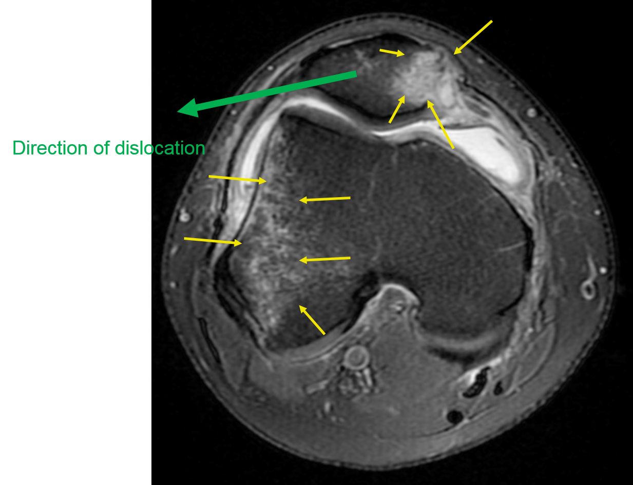
This patient didn’t realise they had dislocated (and relocated) their patella (kneecap) until the MRI scan showed bruising in the bones (white areas arrowed) which occurs when the patella moves sideways over the femur (thigh bone).